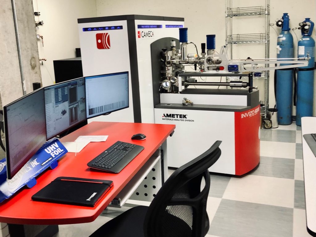
Polytechnique Montréal receives most powerful tomographic atomic probe

Polytechnique Montréal became the first facility to receive the most powerful tomographic atomic probe in North America.
This microscope allows researchers to identify the composition of a sample atom by atom, but also to precisely map the location of each atom.
The potential practical applications are numerous, from developing new treatments for osteoporosis to designing more robust landing gear.
Advertisement
“We’re not just playing Lego with atoms,” said Polytechnique Montréal Professor Oussama Moutanabbir. “If we want to understand the position of atoms, it’s not just for the pleasure of doing it, it’s really to understand the performance of a material and why it will degrade.”
The Invizo 6000 atom probe analyzes the atomic composition of a sample by removing its atoms one by one to generate a three-dimensional image of the object at an unprecedented level of detail.
An integrated mass spectrometre identifies not only the nature of each atom, but also their isotopic form. The tool is so sensitive that it recognizes the smallest atoms, even hydrogen and lithium.
The microscope could enable the development of advanced materials for applications in quantum information technologies: nanoelectronics, optoelectronics, energy conversion and storage, metal alloys for aerospace, bio integrated technologies, and biomaterials.
The technology also makes it possible to consider the design of new generations of semiconductors and quantum materials sensitive to atomic variations and impurities.
Advertisement
Also, the device paves the way to a better understanding of fine structures, like looking inside biological tissues such as bones.
The acquisition of this device was initiated seven years ago and eventually required a partnership between the Université de Montréal, the École de technologie supérieure, McGill University and the Université de Sherbrooke to raise millions of dollars.
The samples that the probe analyzes are about 1,000 times smaller than a human hair. Cut into needle shapes, they are frozen at a temperature of -230 degrees Celsius and subjected to an intense electric field. Pulses from a laser then “lift” the atoms to the surface so that they can be analyzed.
“These intense electric fields make the atoms on the surface loose,” explained Professor Mouttanabir. “Then it takes a few hundred pulses (of laser) to tear the atom away, and once the atom is torn away it will be propelled towards the detector.”
The time it takes for the atom to reach the detector allows researchers to determine its mass and chemical identity. Where the atom hits the detector allows them to calculate where it was on the surface of the sample.
Advertisement
The device has already produced results, such as a meteorite sample that Professor Mouttanabir and his colleagues analyzed.
“We discovered that the meteorite predates the creation of the solar system, so it is more than five billion years old,” the researcher said.
One of Mouttanabir’s colleagues is working, in partnership with industry, on the development of the next generation of X-ray scanners.
To detect cancers as early as possible, he said, you need very efficient detectors, and a key element of that efficiency is the atomic-level homogeneity of the materials used for X-ray scans.
The new device, Mouttanabir said, allows us to “see where the atoms are placed, and their position and distribution will dictate the performance of the detectors.” And if the material used is more uniform, fewer X-rays will be needed to achieve the same result, he said.
Advertisement
“It is also important for everything related to security,” added Professor Mouttanabir. “Soon, in airports, we will have detectors (so efficient) that we will no longer need to empty our bags.”
–This report by La Presse Canadienne was translated by CityNews