Montreal researcher receives $1.2 million to advance glioblastoma research
Posted June 20, 2025 8:19 am.
Last Updated June 20, 2025 8:21 am.
A Montreal researcher recently received a major $1.2 million grant from the Canadian Institutes of Health Research to better understand glioblastoma, a particularly aggressive and lethal form of brain cancer.
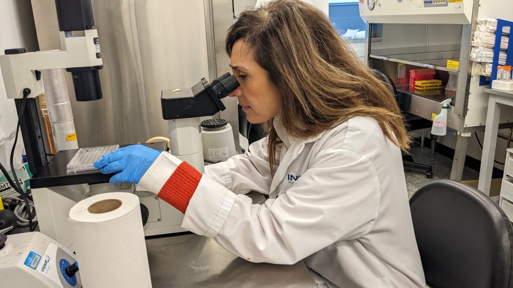
Professor Maya Saleh, from the Armand-Frappier Health Biotechnology Center of the National Institute of Scientific Research, and her colleagues are working to map the immune environment in the brains of patients with glioblastoma.
In particular, they seek to target myeloid cells that contribute to tumor growth and resistance to treatments.
“Despite the initial promise that the immune system could defend us and eliminate cancers if we inject these immunotherapies into patients, we actually see that the pan-cancer response rate is less than 20 per cent,” said Professor Saleh.
“So what interests us are the mechanisms of resistance. We are working on the immunological mechanisms, what combinations of new immunotherapies can we add.”
The average survival time is barely one year after a diagnosis of glioblastoma, a cancer for which treatments have remained virtually unchanged for 40 years.
Treatments for glioblastoma have remained virtually unchanged for 40 years. Standard treatment is surgery, followed by radiotherapy and chemotherapy. However, the tumor recurs in over 90 per cent of cases.
“The first objective (of the grant) is to understand, in fact, the impact of the surgical procedure on tumor progression and relapse,” said Professor Saleh.
It is possible, she added, that the surgery has an inflammatory effect that contributes to tumor relapse, and the grant will also allow for the study of these inflammatory pathways.
Immunotherapy, which primarily uses T lymphocytes, is not very effective because these immune cells are very rarely found in the brain. Instead, monocytes are found there, which can promote tumor growth. Professor Saleh’s work even suggests that surgery could have the perverse and counterproductive effect of stimulating monocyte activity.
The brain is a “very special” organ, the researcher said, and immunotherapy works “at 0 per cent” there.
“When they reach the brain, (some) monocytes become immunosuppressive,” explained Professor Saleh. “So we’re wondering if we can use them as a vehicle to deliver new therapies, if we can modify their character (and) if we can interfere with these immunosuppressive pathways to improve the immune attack of cancer in the brain.”
The problem, she said, is that you can’t completely block monocyte access to the brain, because there will be “cells that are necessary for the immune attack” and only some monocytes change their nature and begin to suppress the immune system in the face of cancer.
“The idea is to understand what signals will differentiate cells (…) and where we could intervene,” explained Professor Saleh. “So when we talk about mapping the immune environment, that’s exactly the goal.”
In each patient, we find “the same immunological signature,” she said, which means that “in that part of the brain, we always have immunosuppression, and in other parts, we will have the good cells. We are trying to map that.”
All clinical trials that have attempted to use immunotherapy for glioblastoma have failed, the researcher added, possibly due to a “lack of understanding of what is happening” in the brain on the immune level.
It is this missing knowledge that Professor Saleh and his colleagues seek to generate.
To validate their work, Professor Saleh’s team is collaborating with Drs. Sami Obaid and Romain Cayrol of the Department of Neurosurgery and Neuropathology at the University of Montreal Hospital. They are analyzing rare biopsies taken from patients who underwent a second operation within 48 hours of the first procedure.
Mass spectrometry is also being used to analyze metabolites present in tissues, in collaboration with Professor Pierre Chaurand of the University of Montreal. This approach makes it possible to examine the impact of glucose, lipids, and other compounds on the tumor environment.
The collected tissues are finally analyzed using cutting-edge technologies such as spatial biology, equipment that INRS is the only institution in Canada to possess. Amadou Barry, an INRS biostatistician, is developing artificial intelligence algorithms to analyze the complex data generated by spatial biology.
“Today, with artificial intelligence, we need to better understand the nature of cancer in order to better treat it,” said Professor Saleh.
For example, she added, it has been found that as the tumor mass grows, the cells in the center begin to lack oxygen, consume glucose and produce lactate.
“And we have a hypothesis that lactate modifies the immune environment by making it more immunosuppressive,” the researcher said. The grant will therefore allow us to study whether lactate receptors could be interesting therapeutic targets.
“There is still a lot to discover, but there is also a lot of progress,” concluded Professor Saleh. “I am optimistic that within a few years we will even have (positive) clinical trials.”
–This report by La Presse Canadienne was translated by CityNews